Head and Neck Surgery
(0618) Characterization of Pleomorphic Adenomas: Determining the Sensitivity and Specificity of MRI as a Diagnostic Tool
Monday, September 30, 2024
12:00 PM - 1:00 PM EDT

Has Audio
Disclosure(s):
Audrey M. Abend, AB: No relevant relationships to disclose.
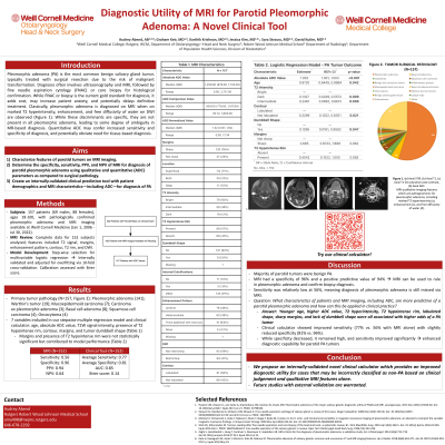
Introduction: Pleomorphic adenoma is the most common benign salivary gland tumor. Historically, the definitive diagnosis before surgical treatment has included ultrasonography with magnetic resonance imaging upon clinical suspicion, followed by fine-needle aspiration cytology and/or core biopsy or surgical resection for histological confirmation. Although highly accurate, the use of FNAC or biopsy incurs additional procedural costs and may potentially delay treatment. Our retrospective study aimed to determine the prevalence at which MRI characterizes and identifies pleomorphic adenomas compared to post-surgical diagnostic pathology.
Methods: We reviewed patient records at Weill Cornell Medicine to determine the consistency between FNAC/biopsy confirmation and positive MRI results for suspected pleomorphic adenoma. A total of 222 patients (93 male, 127 female, and 2 other), ages 18-100, had pathologically proven pleomorphic adenoma and available MRI imaging at Weill Cornell Medicine from January 1, 2006, to July 30, 2022. We examined MRI imaging and utilized T2 signal, margins, enhancement patterns, contour, T2 rim, and diffusion-weighted imaging to characterize tumor morphology.
Results: Pathology reports indicated pleomorphic adenoma (n = 141), Warthin’s tumor (n = 19), mucoepidermoid carcinoma (n = 7), carcinoma ex pleomorphic adenoma (n = 3), basal cell adenoma (n = 8), squamous cell carcinoma (n = 4), and oncocytoma (n = 4). MRI imaging had a sensitivity of 0.39, specificity of 0.95, a positive predictive value of 0.9, and a negative predictive value of 0.54 for the detection of pleomorphic adenoma.
Conclusions: A diagnostic test with high specificity and in turn a high positive predictive value is valuable for clinical decision-making concerning tumor management. For patients with salivary gland tumors, an MRI impression positive for pleomorphic adenoma may be used to rule in pleomorphic adenoma with a high degree of certainty. These results support the prioritization of MRI as a decision-making tool in clinical practice for head and neck surgeons.

Audrey M. Abend, AB
Medical Student
Robert Wood Johnson Medical School
Piscataway, New Jersey, United States- DK
David Kutler, MD
Weill Cornell Medicine Otolaryngology Head and Neck Surgery, United States
